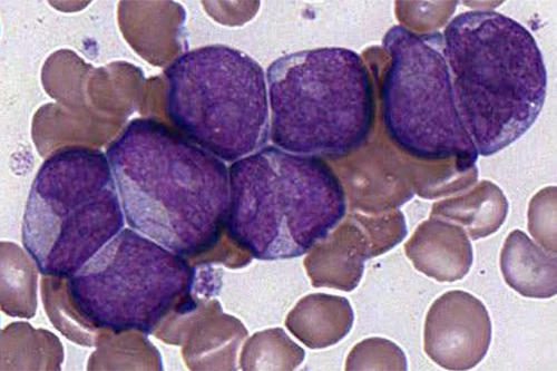
Some ailments can hide their identity from the naked eye, but not the microscope. Through its lens, a trained eye can spot changes in cells and tissues that lead to a quicker diagnosis and often help in managing and treating diseases.
But with COVID-19 driving instruction online everywhere, the college students who will perform these vital services as medical laboratory scientists have lost access to microscope labs and, more vitally, to their stocks of teaching slides. So Stephen Wiesner decided to share his access to online slides.
Wiesner, an associate professor in the University of Minnesota’s Medical Laboratory Science Program, has built a digital database of more than 400 high-resolution pathology microscope slides, covering fields like hematology, bacteriology, parasitology, and body fluids. Through May 31 he is sharing it freely with instructors who train future laboratory scientists, thanks to a licensing option with the U of M Technology Commercialization office.
“Laboratory scientists learn to recognize normal as well as abnormal [blood cell forms and structures],” Wiesner says. “In hematology, abnormally shaped red blood cells aid in the diagnosis of disease in a variety of organ systems, like liver and kidney, and also reflect nutritional deficiencies of iron, vitamin B12, and folic acid. Identification of parasites in blood or other body fluids will help direct appropriate treatment.”
No hospital can do without these services. And by all accounts, Wiesner’s slide database is really catching on. More than 250 educational programs from around the country and internationally have requested access to it.


