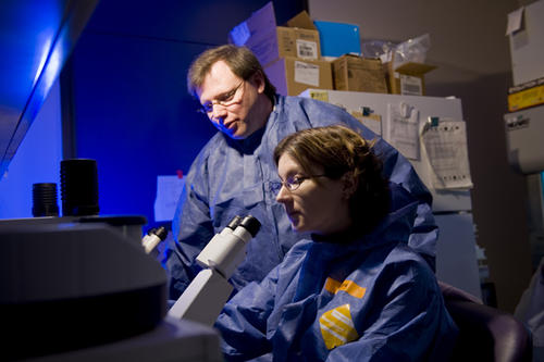
Finding out where a virus replicates inside the body is key to understanding how to stop an infection from spreading. In the case of the first human cancer virus discovered—human T-cell leukemia virus type 1 (HTLV-1)—researchers at the University of Minnesota’s Institute for Molecular Virology (IMV) have discovered a novel pathway for how this distant cousin of the human immunodeficiency virus (HIV) is created from cells.
The study, published in mBio, was the result of looking at live-cell image studies to determine where the major protein needed to create HTLV-1 particles—called Gag—is assembled. These particles are how a virus can spread from one cell to others in the body. The key imaging technique used, called total internal reflection fluorescence (TIRF) microscopy, allowed for precise identification of Gag protein. Researchers discovered it along the periphery of cells.
"With recent and alarming HTLV prevalence studies, there is heightened awareness for increased need for research on this potentially devastating human cancer virus,” said Louis Mansky, PhD, director of the Institute for Molecular Virology, professor in the School of Dentistry and Masonic Cancer Center member. “Uncovering where particle creation occurs will aid in efforts to prevent the spread of this virus."
Just this year, remote parts of central Australia have seen a dramatic rise in HTLV-1 cases. HTLV-1 is transmitted through sexual contact, blood transfusion and from mother to child by breastfeeding. Along with being a carcinogen, the virus can lead to other serious health conditions and cause a chronic progressive disease of the spinal cord. However, most people who are infected never exhibit symptoms.
Mansky and his collaborator, Joachim Mueller, PhD, professor in the School of Physics and Astronomy in the College of Science and Engineering and Masonic Cancer Center member, sought to conduct a careful comparison between how HTLV-1 and HIV-1 particles are formed. The pathway used by HTLV-1 Gag to reach the virus assembly site was distinct from that of HIV-1.
“The use of quantitative fluorescence in live cell imaging experiments allows us to distinguish differences between these viruses,” said Mueller.
"We were surprised by the striking difference observed between HTLV-1 and HIV-1 Gag proteins in our live-cell imaging experiments," said John Eichorst, PhD, senior postdoctoral researcher. “Researchers believed that viruses related to HIV-1 would behave the same, so this difference was quite surprising.”
The next step of this research is to understand the mechanism that determines the pathway used by HTLV-1 Gag to reach the virus assembly site. Blocking this pathway could be an effective means for preventing HTLV-1 transmission and disease.
This research was supported by funding from the National Heart, Lung and Blood Institute, and the National Institutes of Health.
- Categories:
- Science and Technology


