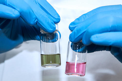
Researchers from the University of Minnesota (UMN) have developed a method to screen and identify harmful or antibiotic-resistant bacteria within one hour using a portable luminometer. Traditional diagnostic methods often require complex equipment and lab work that can take days. The new method uses chemiluminescence, or the emission of light during a chemical reaction. It was developed with the food industry in mind and could also be used in healthcare settings.
In a study published in Advanced Healthcare Materials, researchers from the College of Food, Agricultural and Natural Resource Sciences and the College of Science and Engineering at UMN demonstrated the new technology by analyzing surface swabs and urine samples for the presence of small concentrations of methicillin-resistant Staphylococcus aureus (MRSA), a bacteria that causes more than 11,000 deaths in the U.S. every year.
"A big barrier for microbial detection in the food industry is cost and the inability to detect harmful bacteria in a reasonable time," said John Brockgreitens, a graduate student involved in the study from the Department of Bioproducts and Biosystems Engineering. "We’re trying to develop an inexpensive and rapid way for microbial detection that can be used without needing extensive training."
To screen for microorganisms, green gold in the form of triangular nanoplates was combined with a reducing agent and luminol. This caused a strong chemiluminescent reaction that was stable for as long as 10 minutes. When researchers introduced MRSA and other microorganisms into the combination, they consumed the gold nanoplates, causing the chemiluminescent intensity to decrease proportionally to the microbial concentration. This indicated a presence of microorganisms.
"Rapid microbial detection in less than two hours is not only vital to prevent food poisoning, but also to fight antimicrobial resistance by helping physicians make informed decisions before prescribing antibiotics," said Abdennour Abbas, a professor in the Department of Bioproducts and Biosystems Engineering, who directed the research. "More work is needed to apply this technology to more complex samples such as food and crops, but we’re hopeful that progress will continue in this area.”
Researchers also introduced a new concept called microbial macromolecular shielding to specifically identify MRSA. A polymer specific to MRSA was added to the same sample where it engulfed and surrounded the MRSA bacteria, preventing them from consuming the gold nanoplates. This increased chemiluminescence intensity, indicating the presence of MRSA.
More research is needed before the method can be used in real-world applications, but researchers are eager to make this process faster and easier for industry use.
"In the food industry, items like processed meat, cheese, yogurt and milk have a lot of other competing parts such as proteins and other cells that you need to effectively filter out before you could detect what you’re looking for," Brockgreitens said. "We know our direction is to keep looking at some of these cellular interactions and how to make this whole process either automated or a one-step process.”
This research was funded by the National Science Foundation Award No. 1605191, the University of Minnesota MnDRIVE Global Food Venture, the USDA National Institute of Food and Agriculture Hatch project 1006789, General Mills, the Schwan’s Company Graduate Fellowship, and the Midwest Dairy Association.

- Categories:
- Science and Technology


